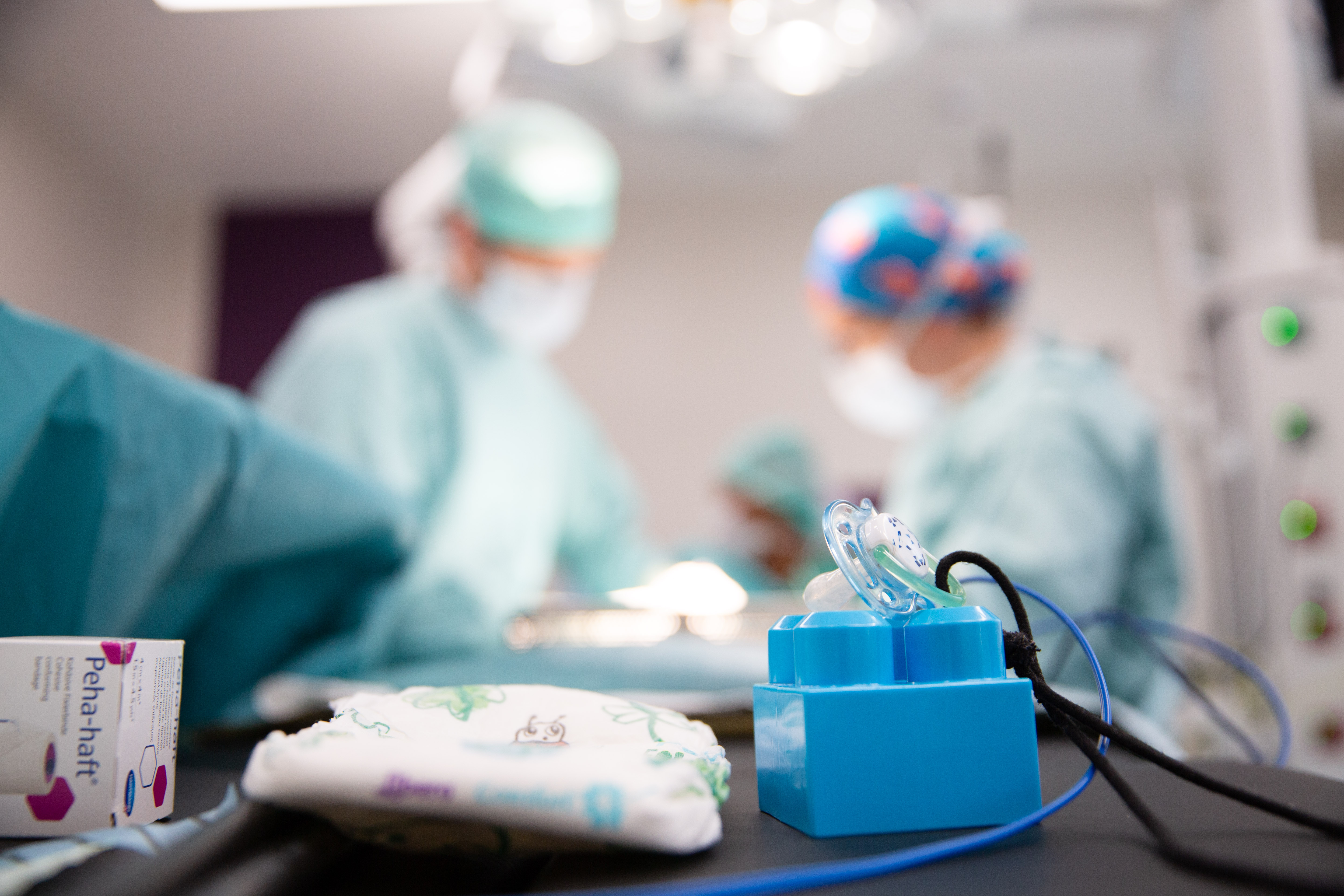

Paediatric Surgery: 40 Years of Pushing the Boundaries of What Is Possible
Over the past forty years, paediatric surgery has undergone profound changes: advances in imaging and anaesthesia, the development of neonatal intensive care, laparoscopy, increasing subspecialisation and, today, augmented surgery.

“Minimally invasive surgery has been one of the major revolutions of recent decades. However, advances in surgery cannot be separated from those made in the areas surrounding it: better diagnosis, improved anaesthesia and better support for children have transformed their care just as much as the surgical procedure itself.”
1986: paediatric surgery enters a new era
The story began well before the construction of the Children’s Hospital. In Brussels, an autonomous paediatric surgery department had existed at Brugmann Hospital since 1923. Over the decades, the discipline became increasingly structured and specialised, notably under the impetus of surgeons such as François Moyson, Willy Grégoir and, later, Frank Collier and Marc-Henri De Laet. In 1974, François Moyson also helped establish the Belgian Association of Pediatric Surgery (BELAPS), demonstrating that paediatric surgery was gradually establishing itself as a discipline in its own right.
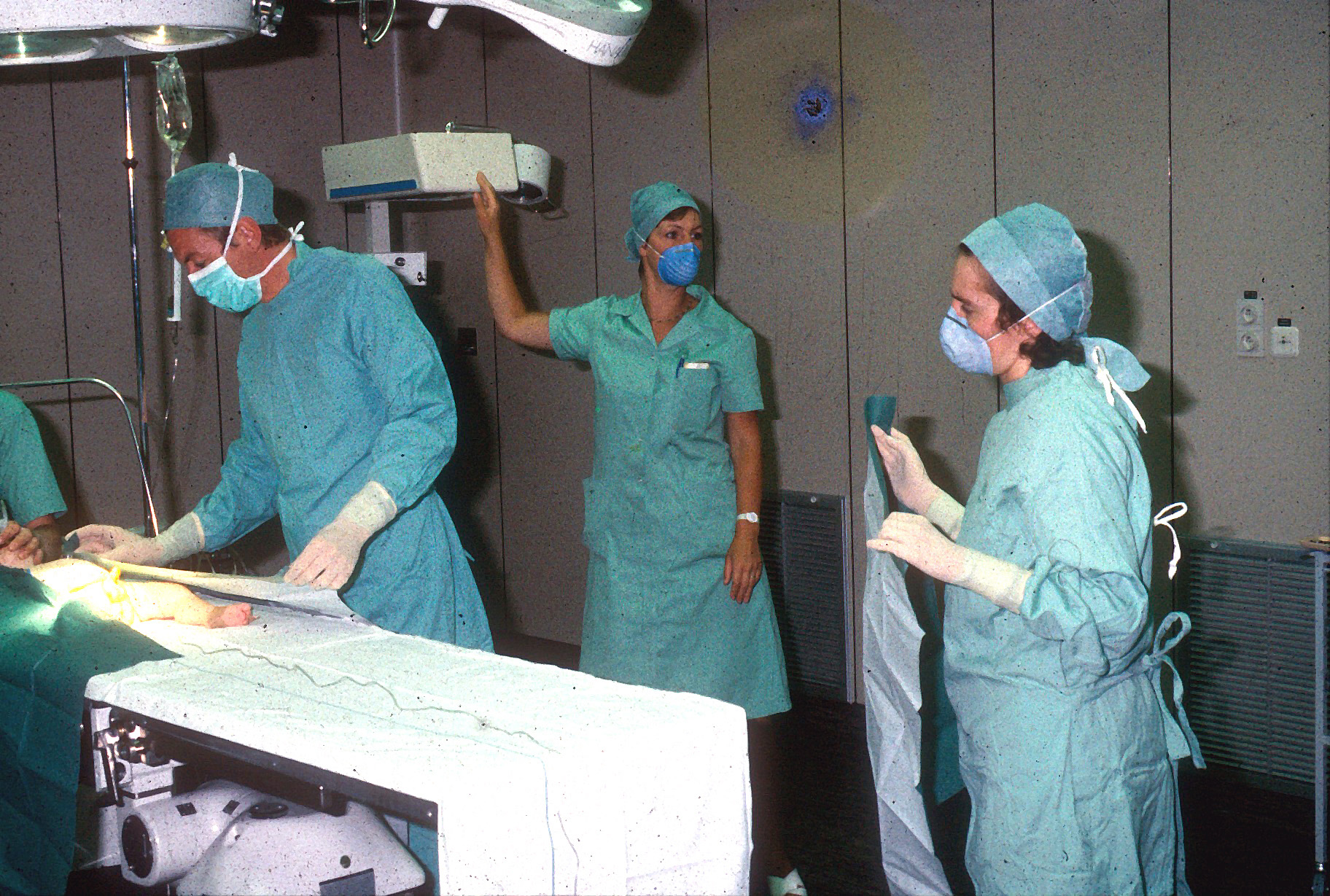
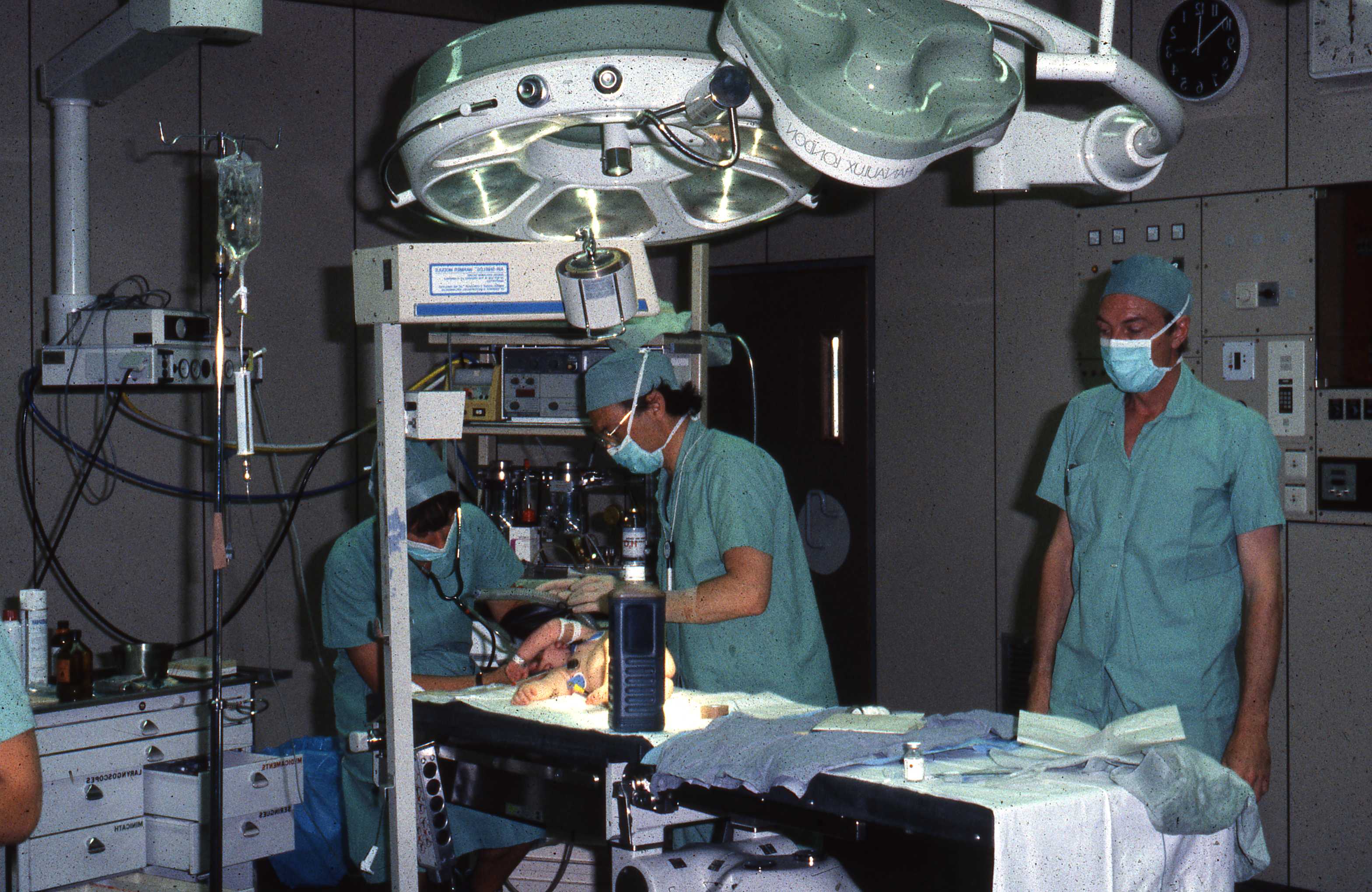
However, 1986 marked a particularly important milestone. With the opening of HUDERF, the various skills required to care for children were brought together in a single paediatric university hospital. Surgeons, paediatricians, anaesthetists, radiologists, neonatologists and intensive care specialists could now work in an environment entirely designed around the child.
This proximity would play an essential role in the decades that followed. Advances in paediatric surgery never depend solely on developments in surgical techniques. Operating on a newborn only a few days old also requires the ability to anaesthetise the infant, provide ventilation, control their temperature, deliver appropriate nutrition and ensure intensive monitoring before and after the procedure.
From the 1980s to the present day: seeing disease before birth
One of the most profound transformations began even before HUDERF opened. From the late 1970s onwards, the development of antenatal ultrasound made it possible to detect during pregnancy certain abnormalities that had previously been diagnosed after birth. Paediatric urology was one of the first fields to be profoundly transformed by this new way of seeing.
Ultrasound technology then advanced rapidly and, in certain situations, was supplemented by foetal MRI. Digestive, urinary or thoracic malformations could be suspected or diagnosed before delivery. The change was considerable: the birth could be planned in an appropriate environment, and the various teams could jointly anticipate the first hours of life.
“For some malformations, care now begins even before birth,” explains Professor Pierre Lingier. This anticipation does not necessarily mean performing more procedures. On the contrary, improved imaging and knowledge also make it possible to better understand the natural course of an abnormality and determine when surgery is needed—and when an intervention is not necessary.
At the same time, paediatric anaesthesia, ventilation, monitoring, parenteral nutrition and neonatal intensive care made considerable advances. Newborns who had previously been extremely difficult to manage can now undergo complex procedures. Neonatal surgery thus illustrates an essential feature of this history: every advance made in the operating theatre also depends on progress achieved around it.


1990s–2000s: the rise of minimally invasive surgery
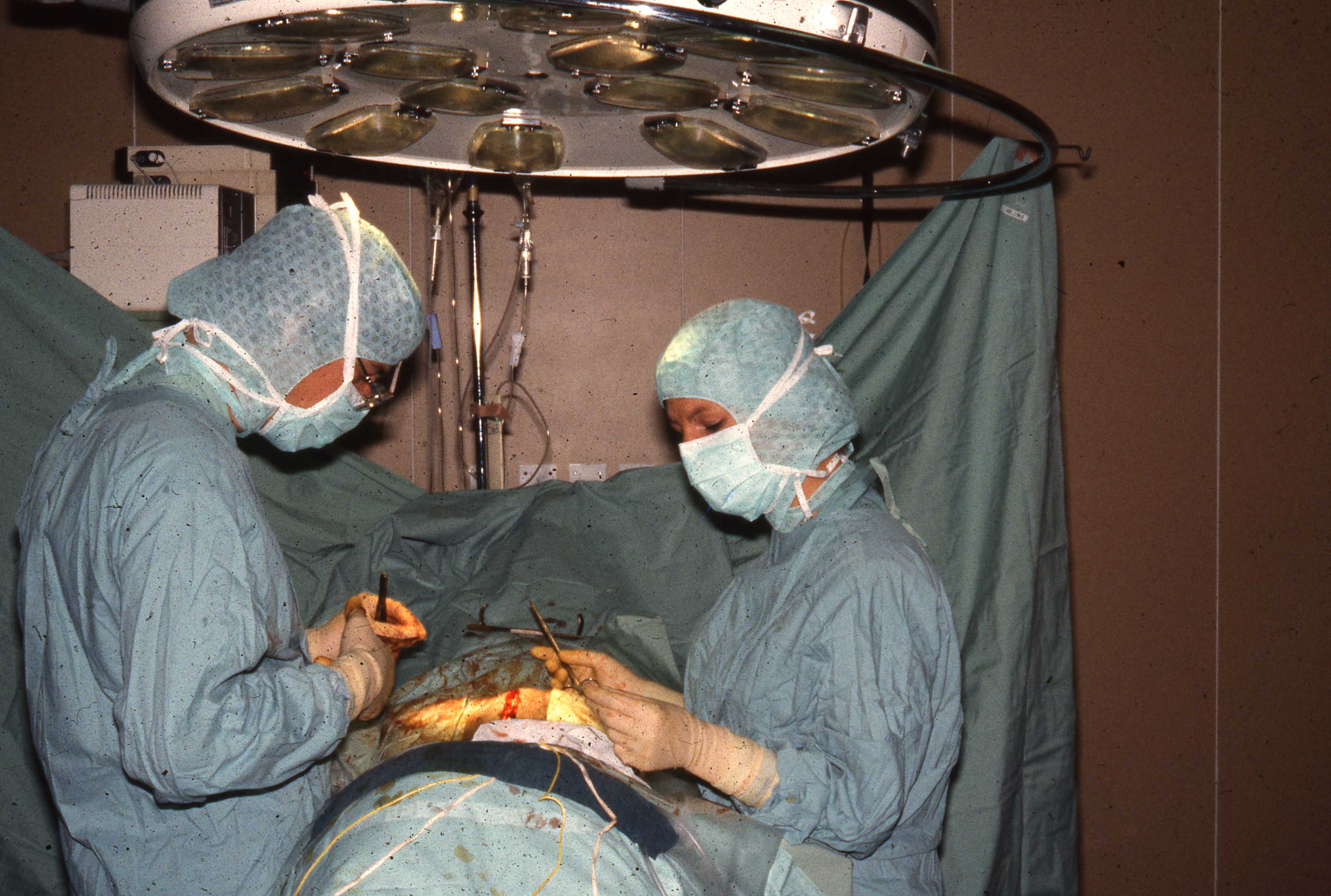
The second major transformation took place in operating theatres. Until the 1980s and the early 1990s, accessing the abdomen or thorax still generally required open surgery. The development of laparoscopy, followed by thoracoscopy, gradually changed this practice.
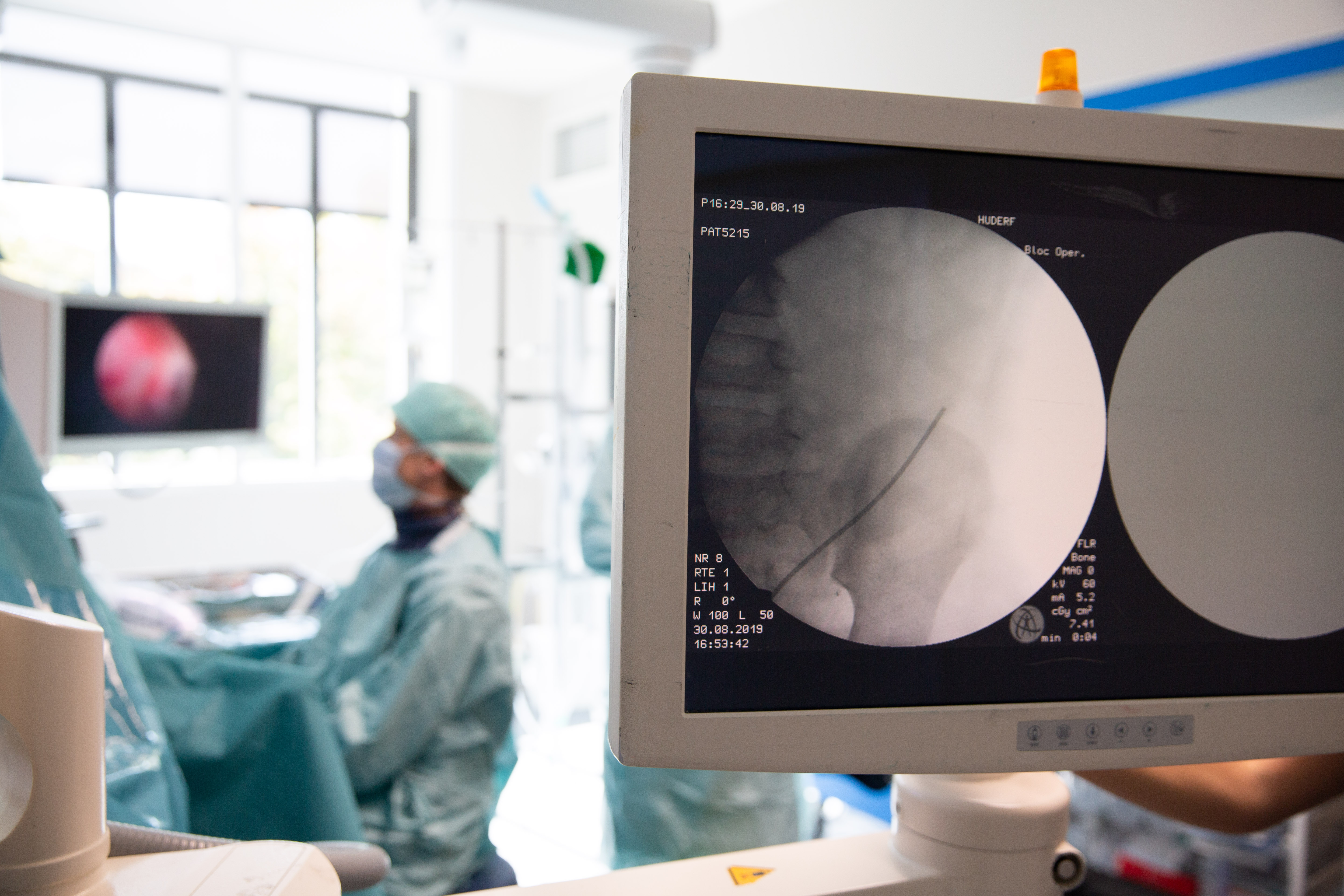
A camera and instruments inserted through small incisions enable the surgeon to operate while viewing a screen. The technique was initially developed in adults, but adapting it to children was far more than simply a matter of scale. The instruments had to be miniaturised, and surgeons had to learn to work in extremely confined spaces, while taking into account the particularly sensitive physiology of infants and newborns.
In Brussels, the first minimally invasive paediatric procedures appeared as early as the beginning of the 1990s. At HUDERF, historical sources place the structured development of laparoscopy and the concept of MIS (Minimally Invasive Surgery) from 2001 onwards.
“For my generation, minimally invasive surgery was probably the greatest technical revolution. Procedures that previously required open surgery can now be performed through incisions measuring just a few millimetres, with, depending on the procedure, less postoperative pain, faster recovery and smaller scars,” explains Professor Pierre Lingier.
This development also went hand in hand with the growth of day surgery and more structured pain management, including after the child returned home. Paediatric surgery therefore changed not only in the way procedures were performed: gradually, the entire care pathway surrounding the child’s operation evolved.
Increasingly specialised surgeons
As techniques progressed, the profession itself evolved. Historically very generalist, paediatric surgery gradually became more specialised: neonatal, digestive, thoracic and hepatobiliary surgery, urology, surgical oncology and renal transplantation each developed their own areas of expertise. For certain rare or complex conditions, the experience of specialised teams became decisive.
HUDERF’s history reflects this development. In urology, the work of Willy Grégoir, particularly on ureterovesical reimplantation, left a lasting mark on the field. Frank Collier subsequently developed paediatric urology at the Children’s Hospital and became Head of the Paediatric Urological Surgery Department in 1999. In digestive surgery, Marc-Henri De Laet, who trained in particular at Great Ormond Street Hospital in London, also contributed to the development of specialised paediatric expertise.
This specialisation, in turn, made it necessary to work more closely within a network. In 2005, Frank Collier became head of the ULB-IRIS interhospital paediatric surgery department, created to improve coordination of paediatric surgical activities among several Brussels hospitals. This approach of sharing expertise continues today within H.U.B, notably between HUDERF, Erasme Hospital and the Jules Bordet Institute for the most complex cases.
The career of Professor Pierre Lingier illustrates this exchange of expertise: after training at ULB and qualifying as a surgeon in 1996, he completed his training in paediatric and neonatal surgery at Necker–Enfants Malades in Paris, before developing a paediatric surgery practice at Erasme and then joining HUDERF. His career reflects the evolution of the discipline: increasingly specialised in terms of expertise, but also increasingly collective in its practice.


2019: an operating theatre designed for modern paediatric surgery
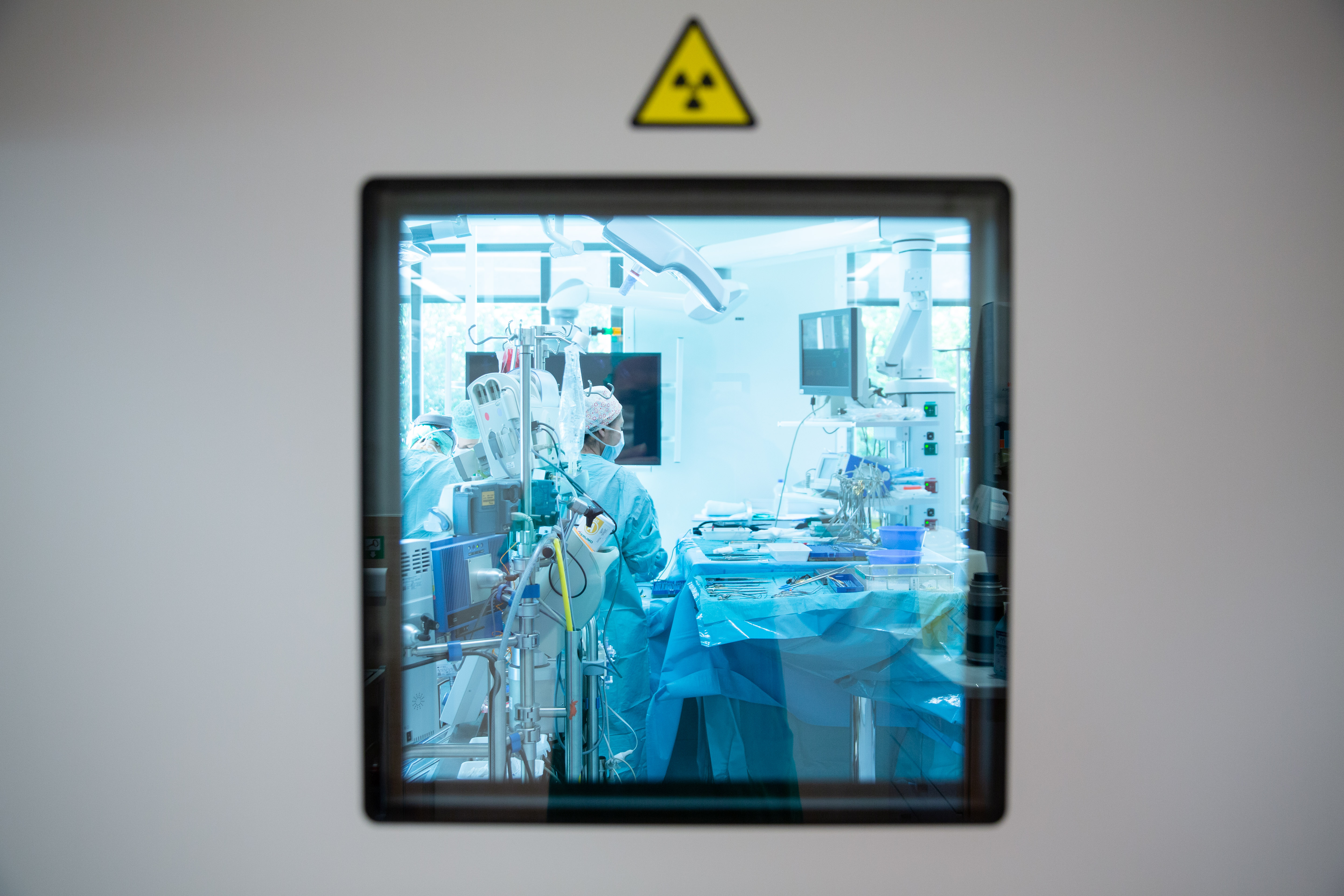
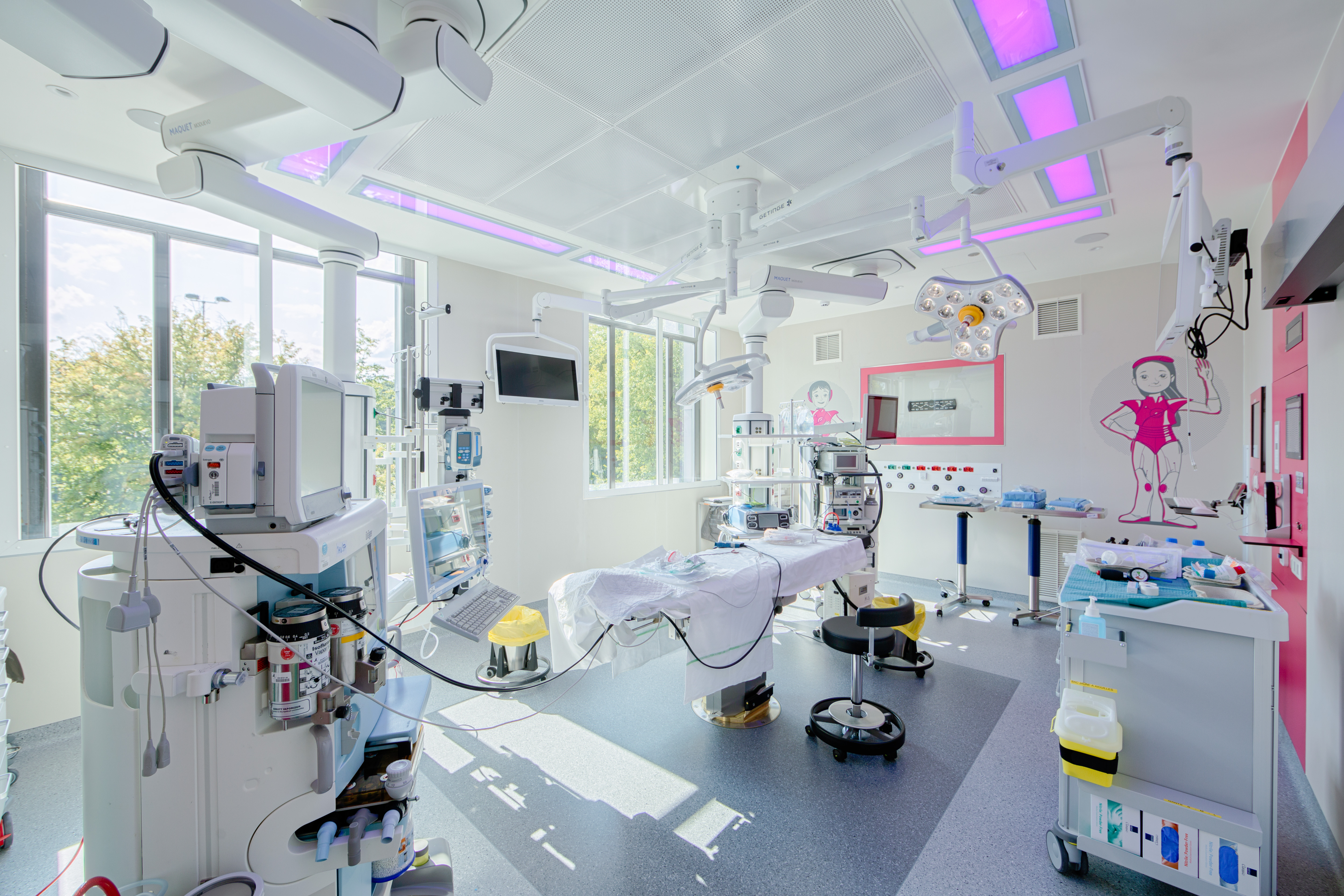
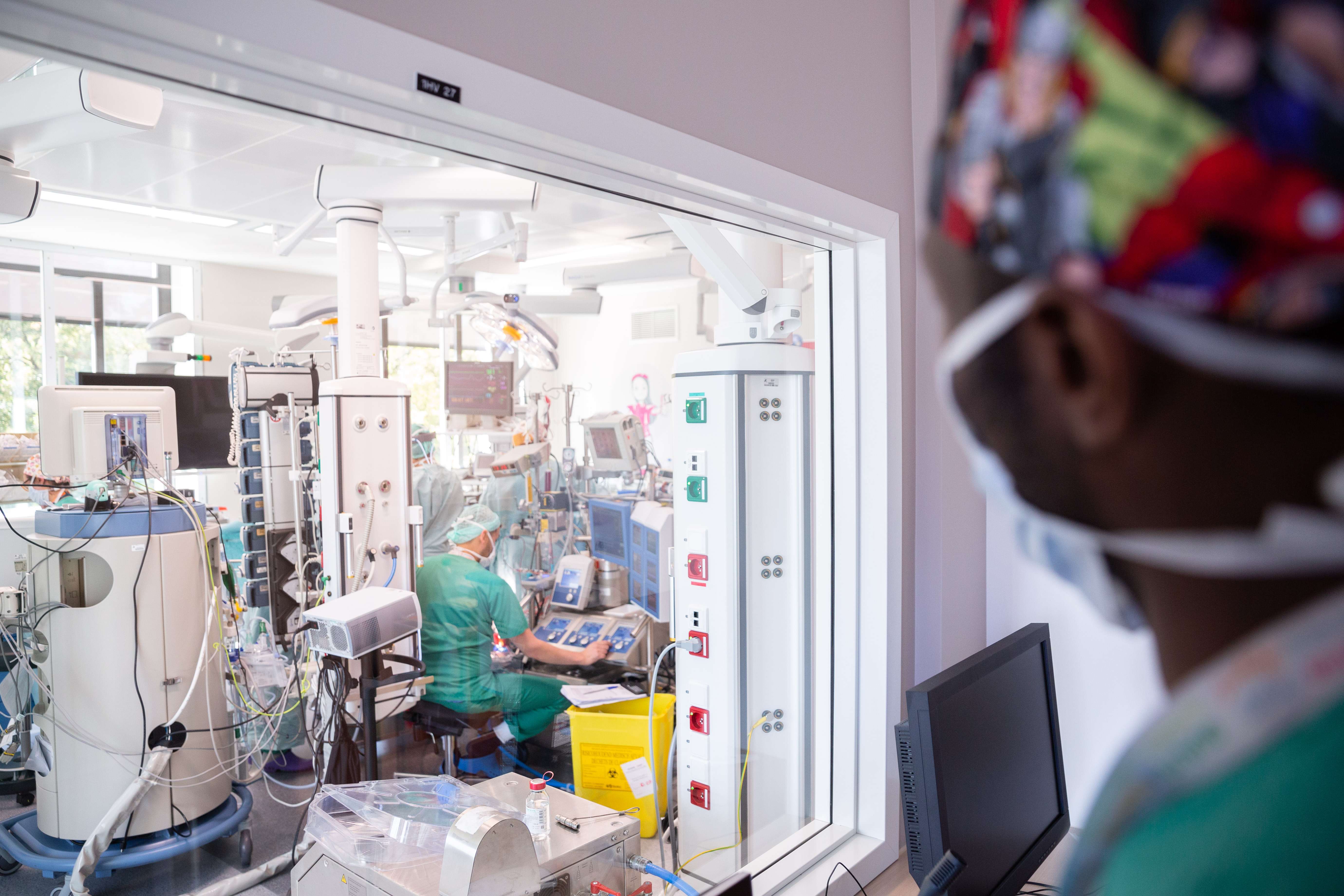
In 2019, the inauguration of the Professor Henri Vis building marked a new milestone for HUDERF, with a modern operating suite adapted to the requirements of paediatric surgery and anaesthesia. At the time, under the leadership of Professor Henri Steyaert, a specialist in minimally invasive paediatric surgery, the department continued to develop these new approaches.
The changes were not limited to the equipment. Over the course of forty years, the entire surgical pathway has evolved: preparation before the procedure, anaesthesia specifically adapted to children, pain prevention and treatment, recovery, the development of day surgery and an earlier return home. Parents’ presence and support for the child have also taken on an increasingly important role. The aim is both to make procedures safer and to reduce their physical and emotional consequences as much as possible.
Today: from minimally invasive surgery to augmented surgery
Following minimally invasive surgery, a new transformation is taking shape. Pierre Lingier readily refers to “augmented surgery”: surgery in which new technologies make it possible to gain a better understanding of the anatomy before the procedure and to guide the surgical movement with ever-greater precision during the operation.
Three-dimensional imaging can already be used to reconstruct certain complex anatomical structures. Fluorescence can help visualise structures that are difficult to distinguish with the naked eye. Robotics opens up prospects for performing highly precise movements in confined spaces, while artificial intelligence could gradually contribute to image analysis and the preparation of procedures. For the surgeon, the aim is not to delegate the surgical movement to technology, but to have new tools available to see better, prepare better and intervene more effectively.
Over the course of forty years, paediatric surgery at HUDERF has therefore undergone several major transformations: the development of antenatal diagnosis, advances in neonatal surgery and anaesthesia, the rise of laparoscopy, the increasing specialisation of teams and, today, the arrival of new imaging and surgical-assistance tools.
The guiding principle, however, has hardly changed: to understand earlier, intervene more precisely and limit the impact of surgery on the child as much as possible. It is this constant pursuit that connects the opening of HUDERF in 1986 with the paediatric surgery that continues to evolve there today.